Paula's
Awsome Web-site
The Microscope
The
first microscope was invented by Zacharias Janssen in 1590. He designed
the first microscope by combining two lenses together. The microscope lets
us see things that anyone wouldn't see with a naked eye. Later
Antony van Leeuwenhoek designed a simple microscope that could magnify
things as much as 270 times. About forty years before Leeuwenhoek other
scientists tried to create compound microscope but they still could not
magnify things more than twenty or thirty times the original size. Luckily
he discovered that if he grinded together two lenses and making sure that
the lighting was just right he created a microscope that magnified things
as much as 270 times. He achieved this and also the images that he had
were so much clearer and brighter than any images of the other microscope.
Robert Hooke later created the compound
microscope by combining two lenses together. A compound microscope is a
microscope that has more than one lenses. The objective lense closest to
the specimen magnifies and projects the image into the body tube of the
microscope where it is further enlarged and projected to the eye. Other
compound microscopes were discovered at this time but Robert Hooke was
the best one. His microscope had three lenses and it closely resembles
the ones used now.
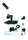
Here is a picture of
a Compound Microscope:
Another very powerful microscope is the Electron
Microscope. It is the most powerful microscope known to man. The Idea was
created by Richard Zsigmondy and Henry Siedentopf. They created a microscope
that lit up the object with an intense beam of light which is very similar
to the Electrone Microscope. Instead the Electron Microscope lights up
the object with and intense beam of electrons. This microscope permits
the observer to view detail far finer then with the best light microscope.
The electron microscope can magnify things as much as 20,000 times then
they can be enlarged up to 1,000,000 times. This allows people to see any
living organism. Molecules are seen through a electron microscope.
The only down side was that the any organism viewed under the Electron
Microscope dies Because of the beam of electrons that shoots through them.
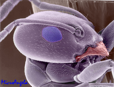
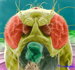
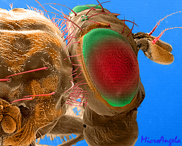
Here are some images
of flies through an Electron Microscope:
There is another type of microscope called
a Dissecting Microscope which is a very low powered microscope to view
organisms that have been dissected. It is very useful to see body
parts with this machine.
 Enough
about school here is what I'm made of.
Enough
about school here is what I'm made of.

My name is Paula Catta. I'm 16 and a Sophmore in High School.
My intrests are dancing, shopping, music, shopping, talking on the
phone, shopping, playing in the band and most important, shopping.
My all time favorite characters are the Smurfs! Remember that 1998
is there 40th Anniversary. Happy Birthday Papasmurf, Smurfet and the rest
of the gang.

My dad, Hugo Catta, is a ham, no not meat but he is into Ham Radio and
thats where he spends most of his time.
His Webpage is mostly
technical and boring
Paula's places to shop. 
http://www.delias.com/main/help/help.htm
l 
http://www.JCrew.com/

http://www.gap.com/onlinestore/gap/

http://www.musicblvd.com/

http://www.guess.com/

 E-mail me @: [email protected]