 GENERAL THERAPY BY RYODORAKU
 LOCAL TREATMENT
 GENERAL THERAPY BY RYODORAKU
- What is Ryodoraku ?
In 1950, Yosio Nakatani found a line, similar to the kidney meridian, that had a series of points in which electroconductivity was higher than the surrounding area, when he was trying to measure skin resistance in general for an edematous patient with nephritis.
Then, he checked this phenomenon carefully in ten kidney disease cases and observed a similar pattern. Healthy subjects did not show this phenomenon that was restricted to kidney disease. Therefore, he named this line, kidney Ryodoraku (ryo is good, do is [electro] conductive, raku is line).
After this, he checked various visceral disorder cases in a similar manner; he recognized that a Ryodoraku corresponding to the classical lung meridian appears in lung disease, a Ryodoraku corresponding to the classical stomach meridian appears in stomach disease, and so on. Thus, Ryodoraku phenomena could be recognized in all classical meridian lines under special abnormal conditions.
Meanwhile, he found that the Ryodoraku matching the "hand's yo-mei large intestine meridian" also sometimes appeared in lung disease. Therefore, the relationship of front-back (positive-negative, Yin-Yang) of the meridians can be proven objectively to some degree.
Since the meridian point is a good electroconductive point, and Ryodoraku which are quite similar to classical meridians actually appear in various pathological conditions, Nakatani concluded that the meridian is a Ryodoraku phenomenon.
Nakatani named meridian points by an easy-to-use method: for example, the meridian (Ryodoraku) of the hand is represented as H, the foot as F. The meridians are numbered consecutively - H1, H2, H3.....etc.
Also, he called H1 the lung Ryodoraku, F6 the stomach Ryodoraku etc., for convenience of comparison with the classical meridian system. The heart envelope meridian (H3) and the triple warmer (H5), which are difficult to understand, were named vascular Ryodoraku and lymph Ryodoraku respectively. In this way, the kidney meridian may well be called the adrenal Ryodoraku, urinary bladder meridian the urinary system Ryodoraku, and the spleen meridian the pancreas Ryodoraku.
Ryodoraku is a pathological phenomenon. Nakatani states that this mechanism can be explained by the viscero-skin sympathetic nerve reflex. The impulses from the viscera radiate to the spinal cord; the reflex zones are then reflected onto the skin surface via the efferent sympathetic nerves and appear as a longitudinal connecting system just like meridian lines (Fig. 1).
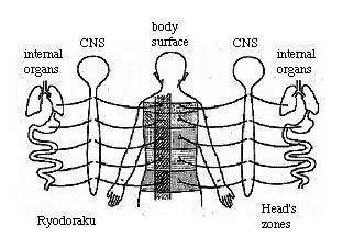 Fig.1 Relationship between Head's zones and Ryodoraku
Sympathetic nerve blocks such as stellate ganglion block for the hand and lumbar sympathetic nerve block for the leg increase considerably the skin resistance in the related area and the reactive electro-permeable points (REPP) or Ryodoraku phenomenon disappear.
Also, the administration of sympathetic nerve stimulants increase electroconductivity in general. Sympathetic blocking decreases the electroconductivity. Accordingly, Ryodoraku may be defined as "the functional route of the excitement in a series of related sympathetic nerves which is caused by visceral disease" or "the linked pathway of the related reactive electro-permeable points".
When adequate stimulation to the reactive electro-permeable point is given, an impulse is induced afferently via the sympathetic nerve and autonomic nerve regulation of the viscera occurs according to Mackenzie's Counter-Concept. As a result of this, electroconductivity at the reactive electro-permeable point is decreased and the symptom is relieved.
Consequently, Nakatani defines Ryodoraku therapy as "measuring skin sympathetic excitability by means of skin elector-resistance and giving stimulation to approach to the normal excitability range through the pathway of nerve reflex.
In oriental medicine, adjusting the basic and essential functions of the whole body in general by considering the physical constitution is called "total functional adjustment". In contrast, treating symptoms only locally called "local treatment".
Balancing fullness and emptiness on meridians is, after all, total functional adjustment. According to Ryodoraku theory, abnormalities on each meridian, or Ryodoraku, can be observed objectively by the measurement of elector-conductivity of certain points on the skin. Then, by stimulating the therapeutic points on the abnormal Ryodoraku, homeostasis ensues and regulates its abnormalities.
Currently, the diagnosis of the disturbance of autonomic nervous system is imprecisely rendered. However, Ryodoraku recognizes each abnormality and can treat them properly. In Ryodoraku theory, an abnormal Ryodoraku has either higher or lower electroconductivity when compared with other Ryodoraku. For this purpose, one calculates the mean value of the Ryodoraku by summerizing the amount of the electroconductive values of the reactive elector-permeable points (REPP) along a Ryodoraku, and dividing by the sum of the total number of reactive elector-permeable points.
In other words, there are twelve elector-permeable points (meridian points) along the LU H1 Ryodoraku . After calibrating the electroconductivity measuring device, one can obtain the electroconductive value for each meridian points and calculate the sum of the twelve electroconductive values. Then, the H1 Ryodoraku average electroconductive value can be obtained by dividing the sum by 12.
Similarly, one can calculate the average value for all of the 24 Ryodoraku. If the average value of H1 Ryodoraku is extremely higher than the others, H1 Ryodoraku is excited (i.e., the H1 sympathetic branch is excited). If extremely lower than the others, it is sedated.
Now, if one gives a stimulation at a given point, a subtle change occurs in the electroconductivity of REPP in the entire body. By observing the changes of the 12 REPP on H1 in this way, it was found that the REPP of H13 showed the average change of the 12 points. Therefore, each time one need not measure the values of 12 points. One can simply measure the H13 electroconductive value and thus observe the average change of H1 electroconductivity. This point was named the "representative measuring point (RMP)" on H1.
In this manner, each RMP was found on the 24 Ryodoraku. Interesting enough, many of these points correspond to the Gen-ketsu (primary meridian point) of each meridian.
3. Electroconductivity Measuring Method of RMP
At first, the neurometer is calibrated as in Fig. 2.

(Neurometer D-401 and DS-207 have been selling by Ryodoraku research Institute Ltd.
...E-mail: [email protected])
Fig. 2 Calibration
procedure to measure RMP
A.
Plug the power cord into
a wall outlet.
B.
Set the
"POWER" switch on then push the operation selector switch
"NEURO"; 110 Volts power supply can be used for detecting. Then turn
the voltage selector switch to "12V".
C.
Insert a cotton swab
saturated with 70% alcohol solution or preferably normal saline into the search
electrode's cup.
D.
Contact the search
electrode to the grip electrode as shown in Fig. 2; turn the current regulator
to the right and adjust the current to 200 micro ampere. Then for measuring,
push the operation selector switch "MEASURE".
- Note: For electroacupuncture treatment, set the operation selector switch to "NEURO". 'Then, this device is in operation with D.C. current.
The position of the patient for measurement as seen in Fig. 3 is ideal, the sitting position with knees bent and feet resting on a small stool or bed and arms extended.
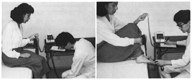
Fig. 3 When trying to measure the patient alone, ideally one positions the Neurometer, patient and practitioner as shown in the picture. One can record the value on the chart with his right hand; and measure the points with his left hand.
 Fig. 4, 5, 6, 7
Location of RMP
Grasp the patient's hand as illustrated. One's thumb and middle finger should point upward. LU-H1 is the point just medial to the thumb. HT-H3 is the point just medial to the middle finger. HC-H2 is in the midline of the wrist joint.
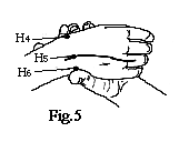 Rotate the wrist and grasp in the same manner as seen
in fig. 4 and 5. SI-H4 and LI-H6 position are similar to LU-H1 and HT-H3.
However, TH-H5 is displaced from the
midline to a point on a line through the center of the ring finger.
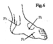 F1: Proximal to 1st metatarsophalangeal Joint
F2: Inferior medial aspect of dorsal prominence of foot
F3: A point on the margin of medial malleolus on a line disecting the heel
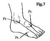 F4: Proximal point on 5th metatarsophalangeal joint
F5: A point on the margin of lateral malleolus and a line through the center of 4th toe
F6: On anterior
margin of dorsal prominence and a line between 2nd and 3rd toes
4. Some notes concerning measurement
During measurement, let the patient grip the electrode but not change the grip hand or grip pressure. The operator's hand should not touch the patient, because often a higher reading results. The operator should not scrub, press, or too often touch the measuring point with operator's finger or search electrode prior to the measurement. Such stimuli change the reading value. Good practice dictates that the search electrode be applied to a point just once.
The search electrode should be placed on each measuring point at a right angle and with equal pressure. Differences in pressure often change the measuring value; thus the amount of pressure should be the same on each point.
Measurement is not ideal immediately following exercise or eating. The patient should rest about ten minutes before measurement. Warming the measuring point should also be avoided.
While the search electrode is on the point, the meter reading has a tendency to rise. Therefore, length of time for the placement of the electrode on the point should be uniform. The absolute value of electroconductivity is less important than the relative value. Some search electrodes are designed with a time limited switch ; a 2.5 second interval insures uniform measuring time.
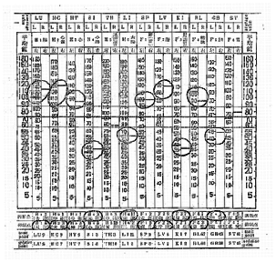 Fig.8 An example of a Ryodoraku chart for a whiplash
injury patient
Electroconductive readings from the
Neurometer on 24 RMP at bilateral hands and feet were recorded. Mean value was
obtained by dividing mathematically the sum of the values of the 24 points (the
line can be roughly drawn). Two fine lines were drawn 0.7cm above or below the
mean value, respectively. This 1.4cm width is the physiological range. When all
readings fall into this range, the autonomic nervous function of the whole body
is well balanced and healthy. The Ryodoraku, the volumes of which are higher or
lower than physiological range, manifest the excitement (fullness) or
inhibition (emptiness) of abnormal Ryodoraku. The therapeutic points according
to the abnormal Ryodoraku are shown at the lower column of the R-chart.
Initially, the patient suffering various symptoms gives quite scattered
readings, as shown here, which, with improvement of the symptoms tend to meet
the physiological range.
Electroconductive readings from the meter (24 representative measuring points in total) for bilateral hands and feet are recorded on the Ryodoraku Chart. Figure 14 is an example of a whiplash injury. The left LU-H1 is 120 micro ampere; and the right LU-H1, is 135 micro ampere, etc.
Secondly, the sum of the 24 points' electro-conductive values divided by 24 gives the mean value. However, clinically, this calculation may be troublesome. Thus, a mean lines roughly can be drawn. Figure 14 shows a line of about 75 micro ampere in mean value. Then, one can draw two lines with a 1.4cm width from the midline (two lines being 0.7cm apart from midline respectively).
This width is the physiological range. When the reading for each Ryodoraku deviates from the physiological range, the is abnormal. Therefore, select the therapeutic point to excite or sedate according to the lower column of the chart. Then, using them, perform general Ryodoraku regulation therapy.
6. The Meaning of the Ryodoraku Chart
1) By investigating many healthy subjects for average electroconductivity, the values shown in Figure 15 were obtained. That is, in each Ryodoraku, the electroconductive readings for healthy subjects differ. According to Nakatani's investigation, the value in summer is higher and in winter lower. Furthermore, the average value differs according to age and sex, and even with the same subject the average value shows some change in each measurement series. Conclusively, Nakatani designed the R-chart so as to be able to record each reading in a healthy condition at the same horizontal level.
 Fig.9 Average Ryodoraku electro-conductive reading for
healthy subjects
2) In the healthy subject, after eating or bowel movement, the electroconductivity or Ryodoraku changes. This allowance is a physiological range; the width of 1.4cm. is a statistical result. When all readings of electroconductivity on each Ryodoraku are within this physiological range, then, the autonomic nervous function is well-balanced and healthy. Often, the charts of athletes display this kind of data. Actually, patients' readings are quite scattered initially; however, with improvement of the symptoms, the scattered points tend to meet the physiological range. The reading is also influenced by various drugs. Tranquilizer or sedative adjust the scattering temporally. Taking a hot bath also makes the scattering well-balanced.
3) When the average value is higher in general, the subject has a hyperstate of the autonomic nervous system. This phenomenon can be observed often in hot weather, youth, or acute diseases. In contrast, the lower average is seen in winter, the aged, or the chronic or fatigued patient. In the latter cases, the response to stimulation therapy is poor.
4) Clinical statistical studies of many cases show what symptoms are indicated by abnormal readings of Ryodoraku. For instance, when KI-F3 Ryodoraku electroconductivity is low (depressed), the patient claims to have less energy or to be impotent. When HC-H2 is higher, (excitation or hyperstate) the patients complain of upperback stiffness in seven out of ten cases. The statistical results may be seen in Table 1.
5) According to Table 1. , Ryodoraku syndromes, one can try a so-called "before-questioning diagnosis." For instance, when HC-H2 and LI-H6 are higher and LV-F2 and KI-F3 lower, one should question the patient as follows: "Have you had upper-back stiffness or impotency recently?" The symptom will be diagnosed with very good probability. This "before-questioning diagnosis" is a very valuable aid in daily practice.
6) This Table was statistically established in a large number of cases who were mostly treated in Internal Medicine. Therefore, if one investigates according to his own medical speciality, there will be some slight variations from this Table . This Table does not either apply to pediatric cases. However, many physicians feel that most adults fit well internationally into the chart. For Americans, too, it was proved that 200 cases tentatively fited this Table . The results are as satisfactory as those for the oriental people.
7) If the measured values are outside of the normal physiological range and Ryodoraku symptoms are not present, it suggests that the patient has a latent symptom or has had an illness in the past.
8) In case eigher the left or right side of a Ryodoraku deviates from the normal physiological range, the disorder is not necessarily located in that particular side. However, when every consecutive measurement indicates the same phenomenon, one can estimate that the particular side has a disorder or abnormality.
9) According to the treatment, deviations from the normal physiological range are gradually diminished. Then, the various symptoms are actually relived. Seasonal change or over-work can affect the patient's condition and cause temporary deviations from the normal range. Recurrence of symptoms takes place again. These findings are valuable in judging the prognosis and therapeutic results, as well as the patient's psychological response.
 LOCAL TREATMENT
7. Electrical Acupuncture (EAP)
Electrical acupuncture is performed with the needle as a stimulative electrode utilizing primarily direct current electricity. The indifferent electrode is held in the hand. A common hypodermic needle may be used to introduce the current ; however, typical electrical acupuncture employs a classical solid needle together with a needle holder as the stimulating electrode. This process is called "electro-needle therapy."
In a localized pain syndrome if sufficient and prompt analgesic effects from mechanical stimulation are desired, continuous or strong stimulation is generally required. (But in some acute cases, very mild stimulation will suffice e.g., mild compression or light massage in the region of a contusion. "In situ" needle technique or miniature ball-patching can also be considered weak stimulation.) Up-and-down manipulation of a needle inserted which is done frequently in daily practice of acupuncture is applied for achieving strong manual stimulation.
When different types of stimulation are used with the needle, e.g., heat or low frequency electric energy, the results differ greatly from simple manual mechanical stimulation. Among the many stimulative techniques, mild direct current to the needle has proven the most adequate and effective for producing analgesia. Some physiological studies have shown that electric energy has an effect 10,000 times greater than that of mechanical energy. Electric energy is, therefore, most effectively used for nerve stimulation.
When applying electrical stimulation to biological subjects, past experience indicates that the stimulative effect reaches supra-maximal level. Further application of electricity does not change the maximum stimulative effect but merely wastes electrical energy and increases the chance of injury. When compared with other sources of stimulative energy, stimulation by mild electrical energy exacts the greatest therapeutic effect.
Direct current may change the peripheral polarization potential in tissues. As such, electrical acupuncture affords strong analgesic effects.